Growth Scans in Pregnancy: Doctor’s Insight
Pregnancy is one of life’s most transformative experiences, both physically and emotionally. As an expectant mother, you’re embarking on a remarkable journey of personal growth that extends far beyond the nine months of gestation. Part of this journey involves understanding the medical assessments that monitor your baby’s development, particularly growth scans. These ultrasound examinations provide crucial insights into your baby’s wellbeing and ensure that everything is progressing as expected.
A growth scan pregnancy ultrasound is a specialized diagnostic tool that measures your baby’s size, weight, and overall development. Unlike the earlier dating scan, a growth scan typically occurs in the third trimester and offers detailed information about fetal growth patterns. Understanding what happens during these scans, why they matter, and what the results mean can help you approach your pregnancy with confidence and informed awareness. This comprehensive guide explores the clinical and personal dimensions of growth scans, helping you navigate this important aspect of prenatal care.
What Is a Growth Scan in Pregnancy?
A growth scan is a specialized ultrasound examination designed to assess your baby’s size, weight, and development during pregnancy. This scan provides measurements of various fetal structures and compares them against established growth charts to determine whether your baby is developing appropriately for gestational age. Medical professionals use growth scans to identify potential complications early, allowing for timely intervention and optimal care planning.
The primary purpose of a growth scan is to evaluate three key aspects: the baby’s biometric measurements (head circumference, femur length, abdominal circumference), the amount of amniotic fluid surrounding the baby, and blood flow patterns through the umbilical cord and placenta. By obtaining these measurements, your healthcare provider gains valuable information about whether your baby is receiving adequate nutrition and oxygen, which are essential for healthy development.
Growth scans represent an important checkpoint in your pregnancy journey, similar to how growth opportunities help us develop in our personal lives. Just as we must assess our progress toward personal goals, medical professionals assess fetal progress through these specialized scans. The information gathered helps create a comprehensive picture of your baby’s health status.
When Are Growth Scans Performed?
Growth scans are typically performed during the third trimester, usually between 28 and 32 weeks of gestation, though they may be recommended at other times depending on individual circumstances. Your healthcare provider might order a growth scan if there are concerns about fetal development, maternal health conditions, or if you’re carrying multiple babies. Some women receive growth scans as routine care, while others may need them only if specific indications arise.
The timing of your growth scan depends on several factors. If you have a history of delivering small babies, if you have gestational diabetes, or if you have high blood pressure, your doctor may recommend an earlier growth scan. Women expecting twins or multiples often have more frequent growth scans to monitor each baby’s development individually. Additionally, if your fundal height measurement (the distance from your pubic bone to the top of your uterus) doesn’t match your gestational age, a growth scan helps clarify whether this is a measurement variation or indicates actual growth concerns.
Understanding the timing of growth scans helps you prepare mentally and physically for the appointment. Like any important assessment, knowing when to expect it allows you to approach the experience with greater calm and readiness.
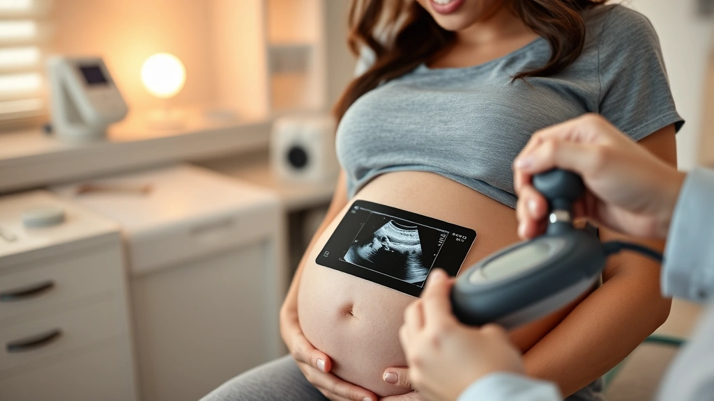
How the Growth Scan Procedure Works
The growth scan procedure is non-invasive and typically takes 20 to 30 minutes, though it may take longer if detailed measurements are needed. You’ll be positioned on an examination table, and a trained sonographer will apply warm gel to your abdomen. The sonographer uses a handheld device called a transducer to transmit sound waves through your skin, creating detailed images of your baby on a monitor.
During the scan, the sonographer takes multiple measurements of your baby’s head, femur bone, abdominal circumference, and other structures. They also assess the amount of amniotic fluid using a measurement called the amniotic fluid index (AFI) or deepest vertical pocket (DVP). Additionally, the sonographer performs Doppler ultrasound studies to evaluate blood flow through the umbilical cord and placenta, which indicates whether your baby is receiving adequate oxygen and nutrients.
The experience is generally comfortable and non-threatening. You’ll be able to see your baby on the monitor, which many women find emotionally meaningful. The sonographer may explain what they’re measuring and may even provide images or videos of your baby. This is an excellent opportunity to ask questions about what you’re seeing and to voice any concerns you might have about your pregnancy.
This assessment mirrors the principle of growth mindset activities, where we actively measure progress and adjust our approach based on feedback. Similarly, growth scans provide concrete data that helps your healthcare team optimize your care plan.

Understanding Growth Scan Measurements
Growth scan measurements are compared against standardized reference charts that account for gestational age. The primary measurements include:
- Head Circumference (HC): Measures the circumference of your baby’s head, which typically correlates with brain development and overall growth.
- Femur Length (FL): Measures the length of the thighbone, which is one of the most reliable indicators of fetal age and growth.
- Abdominal Circumference (AC): Measures the circumference of your baby’s abdomen and reflects liver size, which can indicate nutritional status.
- Estimated Fetal Weight (EFW): Calculated based on the above measurements, this provides an estimate of your baby’s weight compared to other babies at the same gestational age.
Each measurement is plotted on growth charts and expressed as a percentile. For example, if your baby’s weight is at the 50th percentile, it means your baby weighs more than 50% of babies at the same gestational age. Most babies fall between the 10th and 90th percentiles, which is considered normal. Babies below the 10th percentile are considered small for gestational age (SGA), while those above the 90th percentile are considered large for gestational age (LGA).
The Doppler studies measure blood flow velocity in the umbilical artery and middle cerebral artery. Healthy blood flow patterns suggest that your baby is receiving adequate oxygen. Abnormal patterns may indicate that your baby needs closer monitoring or earlier delivery, depending on how advanced your pregnancy is and how concerning the findings are.
Understanding these measurements helps you engage more meaningfully with your healthcare provider. According to research published in the American Journal of Obstetrics and Gynecology, informed patients who understand their ultrasound results report greater satisfaction with their prenatal care experience.
What Normal Growth Looks Like
Normal fetal growth follows a relatively predictable pattern throughout pregnancy. In the first and second trimesters, growth is relatively uniform, and most babies follow similar growth trajectories. However, in the third trimester, growth patterns become more individualized. Some babies are naturally larger or smaller than others, and this variation is completely normal.
A normal growth scan typically shows:
- Measurements that fall within the expected range for gestational age (between the 10th and 90th percentiles)
- Proportionate measurements (head, femur, and abdomen measurements that are relatively consistent with each other)
- Normal amniotic fluid volume (AFI between 8-18 centimeters)
- Healthy blood flow patterns on Doppler studies
- No structural abnormalities or concerning findings
If your growth scan shows normal results, your healthcare provider will likely continue with routine prenatal care and standard delivery planning. Normal growth scans provide reassurance and allow you to focus on preparing for your baby’s arrival. Many women find that a normal growth scan reduces anxiety and allows them to enjoy the final weeks of pregnancy more fully.
It’s important to remember that some variation from “average” measurements is completely normal and doesn’t necessarily indicate a problem. Babies, like all humans, come in different sizes. A baby at the 15th percentile may be perfectly healthy and destined to be a smaller baby, which is simply their natural growth pattern.

Growth Concerns and What They Mean
If your growth scan reveals that your baby is not growing as expected, it’s natural to feel concerned. However, it’s important to understand that abnormal findings don’t always mean something is seriously wrong. Many babies with initial growth concerns deliver healthy babies without complications. Your healthcare provider will discuss what the findings mean and what additional monitoring or interventions might be recommended.
Small for gestational age (SGA) babies are those whose weight falls below the 10th percentile for their gestational age. SGA can occur for several reasons, including maternal factors (smoking, poor nutrition, high blood pressure), placental insufficiency, fetal infections, or genetic factors. Some babies are constitutionally small, meaning they’re naturally smaller but completely healthy. Your doctor will work to determine the cause and develop an appropriate management plan.
Large for gestational age (LGA) babies are those whose weight exceeds the 90th percentile. LGA is often associated with maternal diabetes or obesity. While larger babies can be healthy, they may have increased risks during delivery and immediately after birth. Your healthcare provider may recommend additional monitoring or discuss delivery timing based on your specific situation.
Abnormal Doppler findings suggest that your baby may not be receiving adequate oxygen. This finding requires closer monitoring and may necessitate more frequent scans, non-stress tests, or earlier delivery depending on how advanced your pregnancy is. Research from the Cochrane Library indicates that careful monitoring of pregnancies with concerning Doppler findings improves outcomes.
Reduced amniotic fluid (oligohydramnios) or excessive amniotic fluid (polyhydramnios) can indicate various conditions. Your healthcare provider will assess whether these findings represent true concerns or normal variation. In some cases, increased monitoring is recommended; in others, intervention may be necessary.
Receiving concerning results requires emotional resilience, much like facing any significant challenge. This is where developing a growth mindset becomes invaluable. Viewing these findings as information to guide your care, rather than definitive judgments about your baby’s future, helps you navigate the situation with greater emotional balance.
Emotional Aspects of Growth Monitoring
The emotional experience of undergoing growth scans, particularly when results are unexpected, deserves attention and validation. Pregnancy itself involves significant emotional changes, and the addition of medical monitoring can amplify both excitement and anxiety. Understanding the psychological dimensions of growth scanning helps you navigate this experience with greater self-awareness and support.
Many women experience anxiety before growth scans, wondering whether everything is progressing normally. This is completely natural. The anticipation of receiving information about your baby’s development carries emotional weight. Some women feel relieved after normal results, while others continue to worry despite reassuring findings. This variation in emotional response is normal and doesn’t reflect any deficiency on your part.
If you receive concerning results, you may experience a range of emotions: shock, fear, sadness, or even anger. These reactions are valid responses to information that contradicts your expectations or hopes. Research in International Journal of Mental Health Nursing demonstrates that validation of these emotions, combined with clear information and support, helps pregnant women cope more effectively with stressful prenatal diagnoses.
Several strategies can help you manage the emotional aspects of growth monitoring:
- Seek clear information: Ask your healthcare provider to explain findings in detail. Understanding what the measurements mean reduces uncertainty and fear.
- Bring support: Having your partner, family member, or friend present during scans provides emotional support and helps you process information.
- Connect with others: Talking with other women who’ve had similar experiences can reduce isolation and provide practical perspective.
- Practice self-compassion: Remind yourself that pregnancy concerns are not your fault and that you’re doing everything possible to care for your baby.
- Maintain perspective: Remember that many pregnancies with initial concerns result in healthy births.
Developing emotional resilience during pregnancy mirrors the principles of personal growth. Just as we learn and adapt through challenges, pregnancy challenges can deepen your capacity for strength, patience, and unconditional love. Consider how this experience contributes to your overall growth model as a person and future parent.
If you’re struggling emotionally with pregnancy-related stress or anxiety, don’t hesitate to seek professional mental health support. Many therapists specialize in prenatal mental health and can provide evidence-based strategies for managing anxiety and stress during pregnancy.
FAQ
Is a growth scan safe for my baby?
Yes, growth scans are considered safe. Ultrasound uses sound waves rather than radiation, and no harmful effects have been documented from diagnostic ultrasound use in pregnancy. Thousands of studies have demonstrated the safety of this imaging modality.
Can growth scans accurately predict my baby’s weight at birth?
Growth scans provide estimates of fetal weight, but these estimates have a margin of error, typically plus or minus 10-20% depending on various factors. Actual birth weight may differ from the estimated weight on the scan. The scan’s primary value is tracking growth trends rather than providing exact predictions.
What should I do if my growth scan shows my baby is small?
If your scan shows your baby is small, your healthcare provider will discuss the findings and may recommend additional monitoring, lifestyle modifications, or more frequent scans. Many small babies are born healthy. Your provider will develop an individualized plan based on your specific situation.
Can I request a growth scan if my doctor hasn’t recommended one?
This depends on your healthcare system and insurance coverage. Discuss your concerns with your healthcare provider. If you have specific risk factors or concerns about your baby’s growth, your provider may recommend a scan. Routine scans for all low-risk pregnancies aren’t typically standard practice.
What does it mean if my baby’s measurements are inconsistent?
Inconsistent measurements (where head circumference is at one percentile but abdominal circumference is at a different percentile) can indicate various things, including individual variation, measurement error, or occasionally, specific growth patterns that warrant closer monitoring. Your healthcare provider will interpret these findings in context.
How often are growth scans repeated?
The frequency of growth scans depends on your individual situation. Some women have one scan in the third trimester; others may have multiple scans if concerns arise. Your healthcare provider will determine the appropriate monitoring schedule based on your pregnancy circumstances.